Test Bank for Essentials of Cardiopulmonary Physical Therapy 4th Edition Hillegass ISBN 9780323430548
Course:
Cardiopulmonary Physical Therapy
Institution:
Cardiopulmonary Physical Therapy
Test Bank for Essentials of Cardiopulmonary Physical Therapy 4th Edition Hillegass ISBN 9780323430548 Chapter 1: Anatomy of the Cardiovascular and Pulmonary Systems Test Bank MULTIPLE CHOICE 1. Which of the following chest wall structures is located ...
After purchase, you get:
✅ Instant PDF Download
✅ Verified answer explanations
✅ Refund if not Satisfied
✅ Prepared for 2025/2026 test cycle
Overview
Learners gain valuable practice with question types commonly used on official Bank for Essentials of Cardiopulmonary Physical Therapy 4th Edition Hillegass ISBN 9780323430548 exams. From multiple-choice to case studies, you'll encounter the full range of formats used in actual assessments. This extensive exposure prevents surprises on test day and builds adaptability in your approach. Students often mention how this variety keeps their study sessions interesting and engaging over weeks of preparation.
Who Is This For?
Useful for individuals re-entering assessment practice after a long break, especially those aiming for Bank for Essentials of Cardiopulmonary Physical Therapy 4th Edition Hillegass ISBN 9780323430548 certification. The material helps bridge knowledge gaps efficiently. Many returning students appreciate how it helps them get back up to speed quickly.
Related Keywords
Detailed Study Description
Frequently Asked Questions
Document Information
| Uploaded on: | November 1, 2025 |
| Last updated: | November 17, 2025 |
| Number of pages: | 222 |
| Written in: | 2025/2026 |
| Type: | Exam (elaborations) |
| Contains: | Questions & Answers |
| Tags: | Test Bank for Essentials of Cardiopulmonary Physical Therapy 4th Edition Hillegass ISBN 9780323430548 Chapter 1: Anatomy of the Cardiovascular and Pulmonary Systems Test Bank MULTIPLE CHOICE 1. Which of the following chest wall structures is located level with the second costal cartilage anteriorly and thoracic vertebra T4 and T5 posteriorly? A. Sternal angle B. Jugular notch C. Xiphoid process D. Third costal cartilage ANS: A The sternal angle of the “angle of Louis” is level with the second costal cartilage anteriorly and thoracic vertebrae T4 and T5 posteriorly. PTS: 1 2. Pectus excuvatum is BEST described as: A. Deformity of the sternum caused by trauma B. Caved-in appearance of the chest C. Diminished rib angle anteriorly D. Conical shape of the thoracic cage ANS: B Pectus excuvatum is a common congenital deformity of the anterior wall of the chest, in which several ribs and the sternum grow abnormally; it produces a caved-in or sunken appearance of the chest. PTS: 1 3. The true ribs are BEST defined by which of the following statements? A. Vertebrochondral ribs B. Vertebrosternal ribs C. Ribs 11 and 12 D. Ribs 8, 9, and 10 ANS: B The first seven ribs attach via their costal cartilages to the sternum and are called the true ribs (also known as the vertebrosternal ribs). PTS: 1 |
Seller Information

AdelineJean
User Reviews (0)
Exam (Elaborations)
$17.00
Add to Cart
100% satisfaction guarantee
Refund Upon dissatisfaction
Immediately available after purchase
Available in Both online and PDF
$17.00
| 0 sold
Discover More resources


Inside The Document
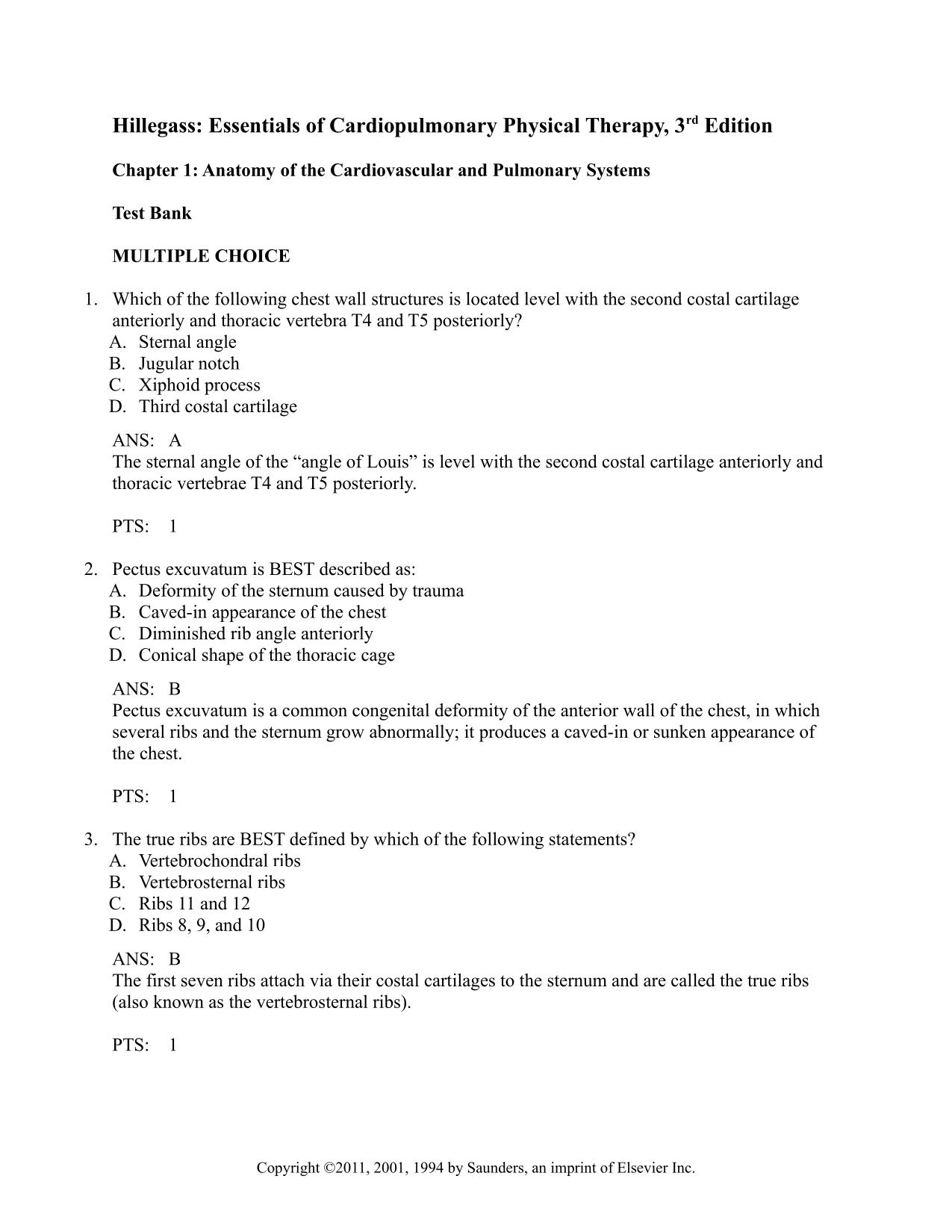
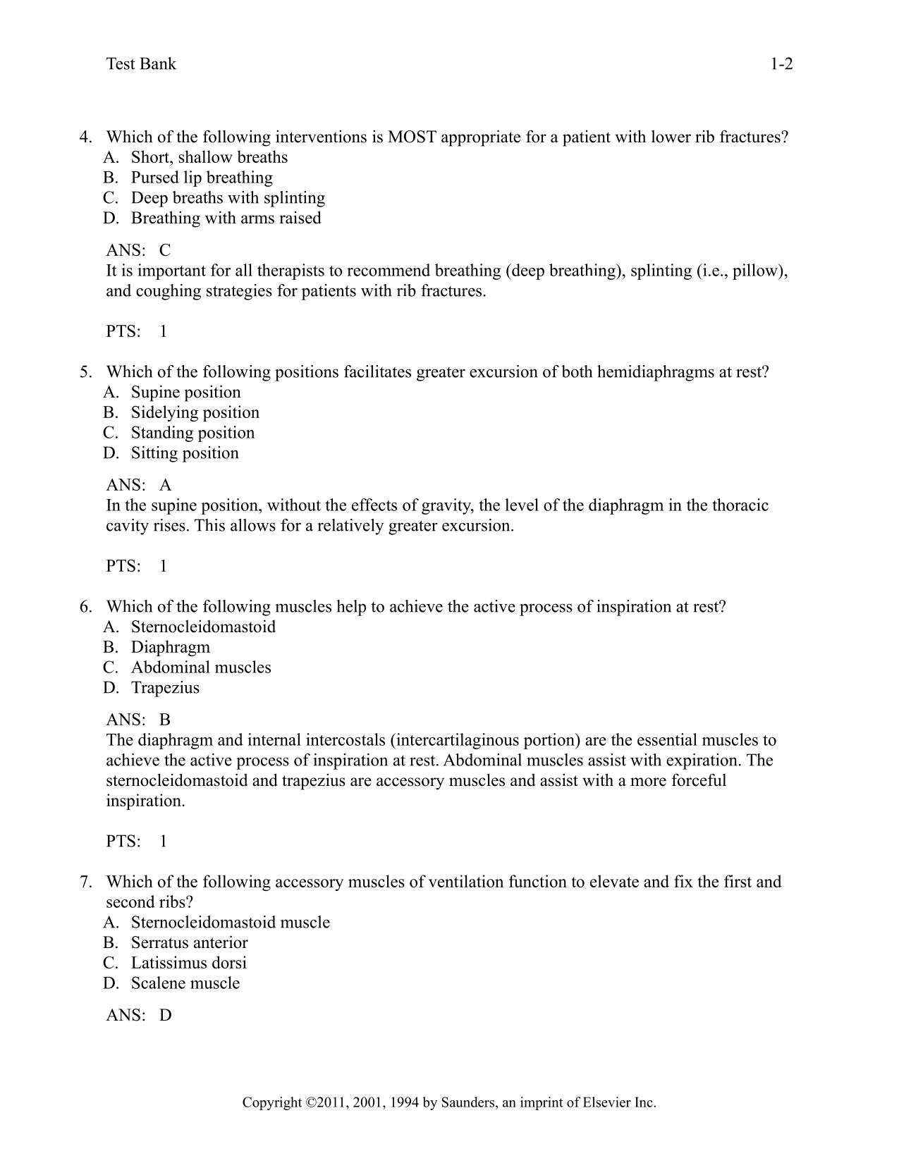
Hillegass: Essentials of Cardiopulmonary Physical Therapy, 3rd Edition Chapter 1: Anatomy of the Cardiovascular and Pulmonary Systems Test Bank MULTIPLE CHOICE 1. Which of the following chest wall structures is located level with the second costal cartilage anteriorly and thoracic vertebra T4 and T5 posteriorly? A. Sternal angle B. Jugular notch C. Xiphoid process D. Third costal cartilage ANS: A The sternal angle of the “angle of Louis” is level with the second costal cartilage anteriorly and thoracic vertebrae T4 and T5 posteriorly. PTS: 1 2. Pectus excuvatum is BEST described as: A. Deformity of the sternum caused by trauma B. Caved-in appearance of the chest C. Diminished rib angle anteriorly D. Conical shape of the thoracic cage ANS: B Pectus excuvatum is a common congenital deformity of the anterior wall of the chest, in which several ribs and the sternum grow abnormally; it produces a caved-in or sunken appearance of the chest. PTS: 1 3. The true ribs are BEST defined by which of the following statements? A. Vertebrochondral ribs B. Vertebrosternal ribs C. Ribs 11 and 12 D. Ribs 8, 9, and 10 ANS: B The first seven ribs attach via their costal cartilages to the sternum and are called the true ribs (also known as the vertebrosternal ribs). PTS: 1 Copyright ©2011, 2001, 1994 by Saunders, an imprint of Elsevier Inc. Test Bank 1-2 4. Which of the following interventions is MOST appropriate for a patient with lower rib fractures? A. Short, shallow breaths B. Pursed lip breathing C. Deep breaths with splinting D. Breathing with arms raised ANS: C It is important for all therapists to recommend breathing (deep breathing), splinting (i.e., pillow), and coughing strategies for patients with rib fractures. PTS: 1 5. Which of the following positions facilitates greater excursion of both hemidiaphragms at rest? A. Supine position B. Sidelying position C. Standing position D. Sitting position ANS: A In the supine position, without the effects of gravity, the level of the diaphragm in the thoracic cavity rises. This allows for a relatively greater excursion. PTS: 1 6. Which of the following muscles help to achieve the active process of inspiration at rest? A. Sternocleidomastoid B. Diaphragm C. Abdominal muscles D. Trapezius ANS: B The diaphragm and internal intercostals (intercartilaginous portion) are the essential muscles to achieve the active process of inspiration at rest. Abdominal muscles assist with expiration. The sternocleidomastoid and trapezius are accessory muscles and assist with a more forceful inspiration. PTS: 1 7. Which of the following accessory muscles of ventilation function to elevate and fix the first and second ribs? A. Sternocleidomastoid muscle B. Serratus anterior C. Latissimus dorsi D. Scalene muscle ANS: D Copyright ©2011, 2001, 1994 by Saunders, an imprint of Elsevier Inc. Test Bank 1-3 The scalene muscles lie deep to the sternocleidomastoid, but may be palpated in the posterior triangle of the neck. These muscles function as a unit to elevate and fix the first and second ribs. The sternocleidomastoid muscle elevates the sternum. PTS: 1 8. When the arms and shoulders are fixed, by leaning on the elbows or grasping onto a table, this muscle can use its insertion as its origin and facilitate an increase in the A-P diameter of the thorax. A. Upper trapezius B. Pectoralis major C. Sternocleidomastoid D. Serratus anterior ANS: B When the insertion and origin of the pectoralis muscle are reversed by leaning on a table to fix the arms, the muscle will pull on the anterior chest wall, lifting the ribs and sternum to increase the A-P diameter of the thoracic cage. PTS: 1 9. The serous fluid within the pleural space serves to provide which of the following functions? A. Create a constant negative pressure B. Assist with venous return of blood to the heart C. Reduce friction between the lungs and thoracic wall D. Serve to allow separation of the pleural layers ANS: C The serous fluid within the pleural space serves to hold the pleural layers together during ventilation and reduce friction between the lungs and the thoracic wall. The space creates the negative pressure to maintain lung inflation, not the fluid itself. PTS: 1 10. Irritation of the phrenic supplied pleura results in which of the following pain referral patterns? A. Thoracic wall B. Abdominal wall C. Mediasternal region D. Lower neck and shoulder ANS: D Irritation of the phrenic supplied pleura can result in referred pain in the lower neck and shoulder, whereas, irritation of the intercostally innervated pleura may result in referral of pain to the thoracic or abdominal wall. PTS: 1 11. An abnormal pleural friction rub on auscultation BEST indicates which of the following? A. Infection with a resultant inflammatory response within the pleura Copyright ©2011, 2001, 1994 by Saunders, an imprint of Elsevier Inc. Test Bank 1-4 B. A buildup of fluid in the pleural space following cardiothoracic surgery C. The presence of blood in the pleural space D. A bacterial infection with resultant pus in the pleural space ANS: A Infection with a resultant inflammatory response within the pleura is termed pleuritis or pleurisy and is best appreciated through the presence of pleural chest pain and an abnormal pleural friction rub on auscultation. A buildup of fluid, blood, or air in the space would result in diminished or absent breath sounds in the area. PTS: 1 12. The presence of four segments (anterior basal, superior basal, lateral basal, posterior basal) BEST describes which of the following lobes? A. Right upper lobe B. Left upper lobe C. Right middle lobe D. Right lower lobe ANS: D The lowermost lobe, the right lower lobe, consist of four segments (anterior basal, superior basal, lateral basal, posterior basal). PTS: 1 13. The physical therapist performs auscultation of the lateral portion of right middle lobe. Which of the following stethoscope locations BEST identifies this lung segment? A. Adjacent to the 5th rib lateral right chest wall B. Adjacent to 3rd–5th rib posterior right chest wall C. Adjacent to the 4th rib lateral right chest wall D. Adjacent to the 8th thoracic vertebra lateral chest wall ANS: A The right middle lobe is subdivided into the lateral and medial lobes. This lobe is the smallest of the three lobes. Its inferior border is adjacent to the fifth rib laterally and sixth rib medially. PTS: 1 14. The BEST reason why a physical therapist should acquire an understanding of the various lobes and segments and their anatomical orientation is which of the following? A. Provide tactile feedback for segmental breathing B. Placement of a stethoscope for auscultation C. Perform appropriate positioning during pulmonary hygiene D. Educate patients on best positioning during coughing ANS: C Copyright ©2011, 2001, 1994 by Saunders, an imprint of Elsevier Inc.
CourseHero & Studypool Unlocks
Get Unlocked CourseHero and Studypool documents files instantly to your email, simply by pasting your link and clicking "Unlock Now". Learn more on how to unlock here.


Gradesity

We are here to help
Questions? Leave a message!
Newsletter
Get notified upon new uploads. Subscribe to our Newsletter.